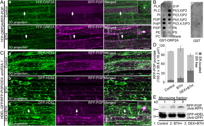
Fig. 8.
PI3P accumulates in the ER membrane during autophagy in ORP2A KD plants. (A) Seven-day-old transgenic YFP-ORP2A/RFP-PI3P seedlings were treated without (a) or with BTH (b) for 6 h, prior to confocal imaging and 3D projection. ΔZ = 0.8 μm, 20 stacks. Arrows indicate examples of colocalized punctae and the enlargement in the box. (Scale bar, 10 μm.) (B) ORP2A binds to multiple phospholipids. Purified GST proteins or recombinant proteins (GST-ORP2A) were subjected to an in vitro lipid-binding assay, followed by immunoblot detection with GST antibodies. (C) Seven-day-old transgenic GFP-HDEL/RFP-PI3P/DEX::amiR2A-1 seedlings were treated without (a) or with BTH (b) for 6 h, followed by confocal imaging and 3D projection. ΔZ = 0.8 μm, 20 stacks. For DEX+BTH treatment (c), DEX pretreatment was performed for 48 h before BTH treatment (c), followed by confocal imaging and 3D projection. Arrows indicate examples of colocalized punctae and enlargement in the box. (Scale bar, 10 μm.) (D) Quantification of the numbers of ER-free and ER-located RFP-PI3P punctae per root section of different treatments shown in C. Error bars indicate ±SD. (E) Immunoblot detection of RFP-PI3P accumulation in GFP-HDEL/RFP-PI3P/DEX::amiR2A-1 plants shown in C. Seven-day-old YFP-ATG1a/DEX::amiR2A-1 seedlings were subjected to treatments as shown in C, followed by isolation of microsomes and subsequent immunoblot detection using RFP or GFP antibodies as indicated. Immunoblot detection of GFP-HDEL using anti-GFP was used as a loading control.

