Fig. 4.
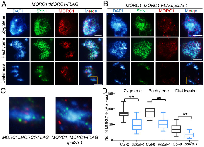
MORC1 foci are significantly reduced in pol2a. (A and B) Anti-FLAG immunostaining showing localization of MORC1-FLAG at zygotene, pachytene, and diakinesis in WT and pol2a-1 background. In WT, MORC1-FLAG tends to form small bodies that are enriched in or near bright chromosome regions (heterochromatin) stained by DAPI at both zygotene and pachytene. In contrast, the MORC1 foci are significantly reduced in pol2a-1. SYN1, a meiosis-specific cohesin subunit, is used as control. (C) Enlarged regions in the yellow boxes from A and B. (D) Comparison of MORC1-FLAG foci counts in Col-0 and pol2a-1 at zygotene, pachytene, and diakinesis. Error bars indicate confidence interval. **, P < 0.01; two-tailed Student’s t test. Scale bars, 5 μm.

