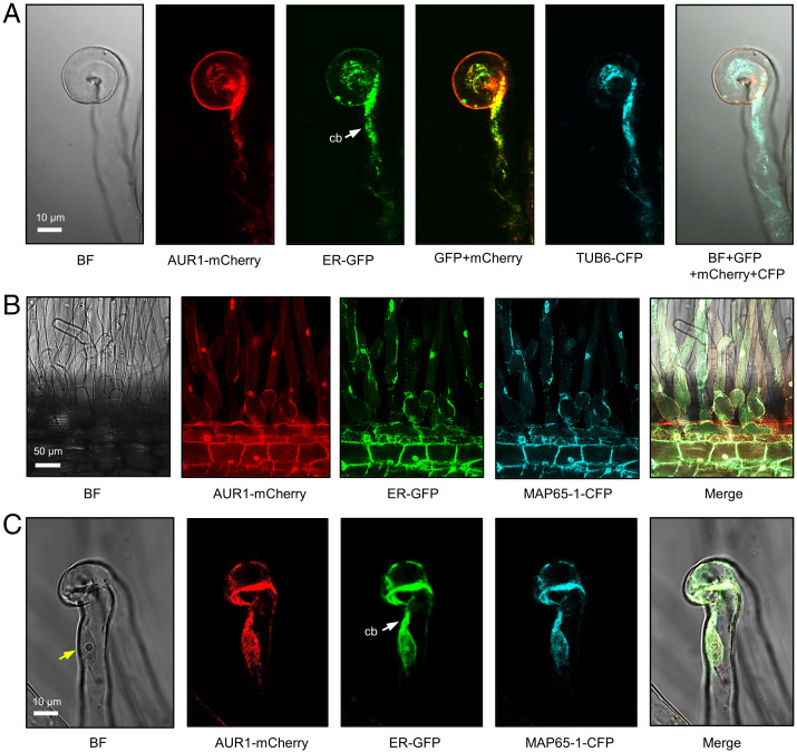
Fig. 3.
AUR1, TUB6, and MAP65-1 localize to the ER in rhizobia-infected root hairs. (A) Confocal images of a curled root hair showing subcellular localization of AUR1-mCherry, ER-GFP, and TUB6-CFP driven by constitutive promoters in M. truncatula A2 hairy roots. (B and C) Confocal images of AUR1-mCherry, ER-GFP, and MAP65-1-CFP in M. truncatula hairy roots. The white arrow indicates the cytoplasmic bridge (cb). The yellow arrow indicates the nucleus of the root hair. Scale bars, 10 μm (A and C) or 50 μm (B). Images were captured in A2 plants, which contain 35Spro:GFP-HDEL at 5 dpi with S. meliloti Rm2011. Experiments were carried out three times with similar results. BF, bright field.

