Fig. 5.
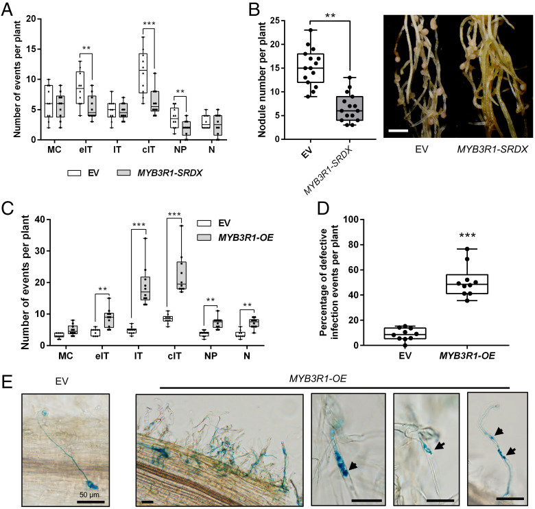
MYB3R1 contributes to nodule symbiosis. (A) Rhizobial infection events, formation of nodule primordia, and nodules in the EV (empty vector) control and MYB3R1-SRDX plants at 7 dpi. MC, rhizobia microcolony; eIT, elongating infection thread in root hair; IT, infection thread that fully traversed the root hair; cIT, infection thread that traversed the root hair and ramified into the underlying cortical cell; NP, nodule primordium; N, nodule. (B) Expression of MYB3R1-SRDX resulted in reduced nodulation at 21 dpi. Representative hairy roots expressing EV or MYB3R1-SRDX are shown in the Right panel. Scale bar, 2 mm. (C) Quantification of infection events including normal and aberrant infections, nodule primordia, and nodules in the EV control and MYB3R1-OE plants at 7dpi. (D) Frequency of defective infection events in EV control and MYB3R1-OE plants. (E) A normal infection event in the EV control plants (Left panel). Typical defective infection events found in MYB3R1-OE plants. A cluster of infection events with examples of abnormal infection events is shown. Arrowheads indicate defective infection threads in root hairs. Scale bars, 50 μm. Boxes show the first quartile, median, and third quartile; whiskers show minimum and maximum values; dots show data points. Means were compared using Student’s t test. **P < 0.01, ***P < 0.001. Experiments were carried out twice with similar results.

