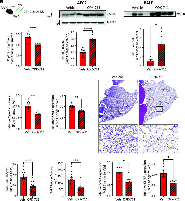
Fig. 7.
IRE1α inhibition reduces the reprogrammed AEC2 cell state and AEC2 driven alveolitis in vivo. (A) Schematic for evaluating in vivo IRE1α inhibition in SftpcC121G mice using OPK-711 (20 mg/kg/day) or vehicle treatment. (B) Xbp1 splicing ratio as assessed by qRT-PCR at day 7 (n = 6 per group) referenced against SftpcWT. (C) Western blotting (Top) of AEC2 lysate for SP-B and densitometry analysis (Bottom) show increased mSP-B in AEC2 cells with OPK-711 IRE1α inhibition (see SI Appendix, Fig. S12C for additional Western blotting for densitometry). (D) Western blotting of BALF large-aggregate surfactant fractions (Top) and densitometry (Bottom) for mSP-B showed increased content in alveolar compartment with OPK-711 IRE1α inhibition. (E) Cldn4 (Left) and Krt8 (Right) expression determined by qRT-PCR of AEC2 cells at day 7 (n = 6 per group). (F) Representative H&E staining of lung sections shows reduced alveolitis in OPK-711-treated lungs (Top: 4× magnification scale bars = 1,000 μM, Bottom: 20× magnification, scale bars = 100 μM). (G and H) BALF granulocytes (G) quantified by manual counting of Giemsa-stained cytospins and expressed as a percentage of total BALF cells and BALF total protein (H) as assessed by Lowry assay in vehicle and OPK-711-treated SftpcC121G mice (n = 11 per group). (I) Ccl11 (Left) and Ccl17 (Right) expression determined by qRT-PCR of AEC2 cells at day 7 (n = 6 per group). *p < 0.05, **p < 0.005, ***p < 0.0005 by two-way t test.

