Fig. 2.
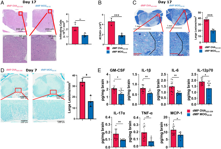
dMP MOG treatment in advanced EAE reduced demyelination, immune infiltration, and inflammatory cytokine levels in the CNS. Mice were induced with EAE and treated at a score of 3, as indicated in Fig. 1B with a second injection 3 d later. (A) Representative images of hematoxylin and eosin–stained sections, and quantification of infiltrating immune cells in the lumbar spinal cord on day 17 posttreatment. Cells were counted and normalized to the area to calculate cells/mm2. n = 4/group. (B) Quantification of CD45+ CNS infiltrates by flow cytometry on day 17 posttreatment. n = 3 to 4/group. (C) Representative images, and quantification of demyelination using luxol fast blue staining in the lumbar spinal cord on day 17 posttreatment. Lesions were counted and those with an area >400 µm2 were classified as a large. n = 3 to 4/group. (D) Representative images and quantification of demyelination using luxol fast blue staining in the lumbar spinal cord on day 7 posttreatment. n = 3. (E) Luminex xMAP technology for multiplexed quantification of mouse cytokines, chemokines, and growth factors from complete brain homogenate at a concentration of 4 mg/mL on day 7 post treatment. n = 8 to 11. d, days post treatment; *P < 0.05; **P < 0.01.

