Fig. 4.
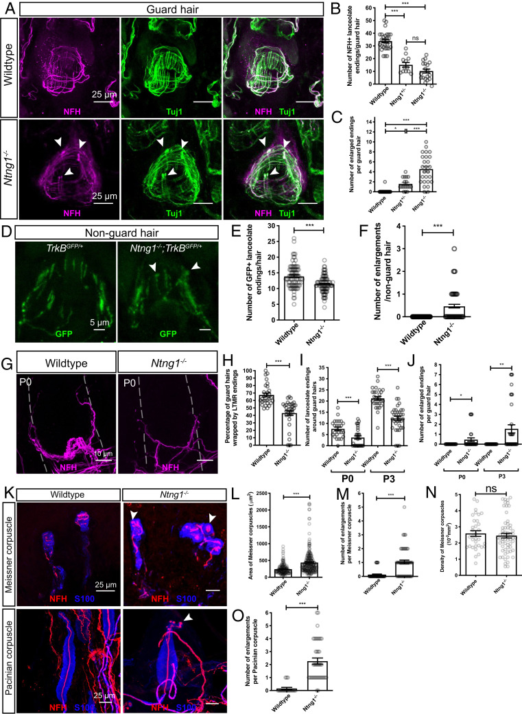
Netrin-G1 regulates the formation of lanceolate endings, Meissner corpuscles, and Pacinian corpuscles. (A) Wholemount immunostaining images of guard hairs in back hairy skin of adult wild-type and Ntng1−/− animals. Aβ RA-LTMRs lanceolate endings are marked by NFH (magenta) and Tuj1 (green) labeling. (B and C) Quantification of the number of NFH+ lanceolate endings (B) and the number of enlarged endings (C) per guard hair for Aβ RA-LTMRs in wild-type (B: 29 hair follicles from five animals; C: 42 hair follicles from seven animals), Ntng1+/− (B: 12 hair follicles from two animals; C: 17 hair follicles from two animals) and Ntng1−/− (B: 19 hair follicles from three animals; C: 26 hair follicles from three animals) animals, showing fewer Aβ RA-LTMR lanceolate endings in Ntng1+/− and Ntng1−/− animals. Each dot represents a single hair follicle. One-way ANOVA test. (D) Wholemount immunostaining images of nonguard hairs in back hairy skin in TrkBGFP/+ and Ntng1−/−; TrkBGFP/+ animals. Aδ-LTMRs lanceolate endings are marked by GFP labeling. (E and F) Quantification of the number of GFP+ lanceolate endings (E) and the number of enlarged endings (F) in TrkBGFP/+ (90 hair follicles from three animals) and Ntng1−/−; TrkBGFP/+ (95 hair follicles from three animals) animals, showing similar deficits in lanceolate ending formation with nonguard hairs. Student’s unpaired t test. (G) Wholemount immunostaining images of guard hairs in back hairy skin of P0 wild-type and Ntng1−/− animals. (H) Quantification of the percentages of guard hairs wrapped by NFH+ lanceolate endings at P0 in wild-type (29 hair follicles from three animals) and Ntng1−/− (33 hair follicles from three animals) animals. Student’s unpaired t test. (I and J) Quantification of the number of NFH+ lanceolate endings (I) and the number of enlarged endings (J) per guard hair in back hairy skin for Aβ RA-LTMRs in wild-type (25 hair follicles from three animals for P0; 28 hair follicles from five animals for P3) and Ntng1−/− (31 hair follicles from three animals for P0; 25 hair follicles from five animals for P3) animals. Student’s unpaired t test. (K) Representative IHC images of forepaw glabrous skin sections and Pacinian corpuscles. Meissner corpuscles and Pacinian corpuscles are labeled by S100 (blue) for visualizing lamellar cells and NFH (red) for visualizing Aβ RA-LTMRs. Arrowheads point to axonal enlargements. (L–N) Quantification of the area (L), number of enlargements (M), and density (N) of Meissner corpuscles in the epidermis of wild-type (44 skin sections from four animals) and Ntng1−/− (60 skin sections from four animals) mice. Each dot represents a single skin section (O). Student’s unpaired t test. (O) Quantification of the number of enlargements per Pacinian corpuscle from wild-type (19 corpuscles from three animals) and Ntng1−/− (41 corpuscles from three animals) mice. Student’s unpaired t test. Each dot represents a single Pacinian corpuscle. ns, not significant, *P < 0.05, **P < 0.01, ***P < 0.001.

