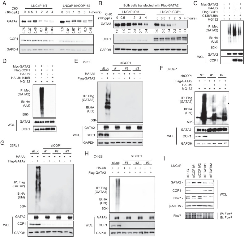
Fig. 2.
COP1 is an E3 ubiquitin ligase for GATA2. (A) LNCaP cells with 2 µg/mL Dox-induced knockdown of COP1 (ishCOP1#2) versus nontargeting control (iNT) were treated with 10 ng/µL chlorhexidine (CHX) for 0–4 h. (B) Flag-tagged GATA2 was transfected into LNCaP cells with 0.5 µg/mL Dox-induced ectopic expression of COP1 (iCOP1) versus control (iCtrl). After 48 h, the cells were treated with 10 µM MG132 for 2 h. MG132 was then removed, and cells were treated with 10 ng/µL CHX for 0–4 h. Western blots were quantified using Image J. (C) Ubiquitination assay. HEK293T cells were transfected with Myc-tagged GATA2 (Myc-GATA2), HA-tagged ubiquitin (HA-Ubi), and Flag-tagged COP1 (Flag-COP1) versus an E3 ligase-dead COP1 mutant (Flag-C136/139A). After 48 h, the cells were treated with 10 μM MG132 for 6 h before preparing cell lysate for the indicated immunoprecipitation (IP) and Western blot (IB) analyses; WCL, whole cell lysate. (D) HEK293T cells were transfected with Myc-GATA2, Flag-COP1, or HA-Ubi versus HA-Ubi K48R mutant. After 48 h, the cells were treated with 10 μM MG132 for 6 h before preparing cell lysate for IP and IB analysis. (E) HEK293T cells were transfected with Flag-tagged GATA2 (Flag-GATA2), HA-Ubi, and three different siRNAs against COP1 (siCOP1) versus control (siLuc). After 72 h, the cells were treated with 10 μM MG132 for 6 h before preparing cell lysate for IP and IB analysis. (F) LNCaP cells with COP1 knockdown (shCOP1 #1 and #2) versus control (NT) were transfected with Flag-GATA2 and HA-Ubi. After 48 h, the cells were treated with 10 μM MG132 for 4 h before preparing cell lysate for IP and IB analysis. (G) 22Rv1 and (H) C4-2B cells were transfected with three different siRNAs against COP1 (siCOP1) versus control (siLuc). After 24 h, the cells were transfected with FLAG-tagged GATA2 (Flag-GATA2) or HA-Ubi. After 48 h, the cells were treated with 10 μM MG132 for 4 h before preparing cell lysate for IP and IB analysis. (I) LNCaP cells were transfected with siLuc control or two independent siRNAs against COP1 (siCOP1) or FBW7 (siFBW7). After 72 h, the cell lysates were prepared for IB. IP with anti-Fbw7 followed by IB with anti-Fbw7 was used to further confirm knockdown of Fbw7. All data are representative of at least three independent experiments and are presented as Western blots.

