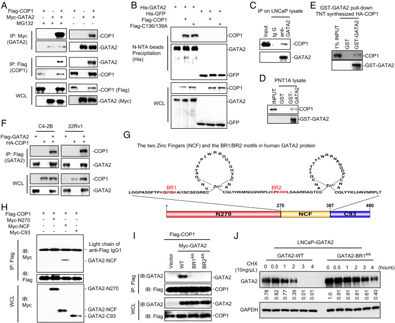
Fig. 3.
The BR1/BR2 motifs of GATA2 are essential for COP1 binding. (A) HEK293T cells were transfected with Myc-tagged GATA2 (Myc-GATA2) and Flag-tagged COP1 (Flag-COP1). After 48 h, cells were treated with 10 μM MG132 versus vehicle control for 6 h before preparing cell lysate for IP using anti-Myc antibody (GATA2) or anti-Flag antibody (COP1) and for IB analysis. (B) HEK293T cells were transfected with His-tagged GATA2 (His-GATA2) versus control His-tagged enhanced green fluorescent protein (eGFP) and Flag-COP1 versus an E3 ligase-dead COP1 mutant (Flag-C136/139A). After 48 h, the cells were treated with 10 μM MG132 for 6 h before preparing cell lysate for His-tagged protein precipitation using Ni-NTA beads and IB analysis. (C) LNCaP cells were treated with 10 μM MG132 for 4 h before preparing cell lysate for the indicated IP and IB analysis. (D) GST-tagged GATA2 (GST-GATA2) protein and GST protein (as control) were overexpressed in E. coli and purified and conjugated onto glutathione Sepharose beads. The beads were then used in the pull-down assay for GATA2 binding with the endogenous COP1 in the PNT1A cell lysate. (E) COP1 protein was synthesized using the TNT In Vitro Kit. GST-GATA2 protein-conjugated beads were used in the pull-down assay for GATA2 binding with the COP1 synthesized in vitro. (F) C4-2B and 22Rv1 cells were transfected with Flag-tagged GATA2 (Flag-GATA2) and HA-tagged COP1 (HA-COP1). After 48 h, cells were treated with 10 μM MG132 for 4 h before preparing cell lysate for IP using anti-FLAG antibody (GATA2) and IB analysis. (G) Illustration of the two zinc finger domains in human GATA2. The basic K/R residues in the BR1 and BR2 motifs are highlighted in red. (H) HEK293T cells were transfected with Flag-COP1 and Myc-tagged truncates of GATA2, including N270 (N-terminal 1–270 aa of GATA2), NCF (N- and C-terminal zinc finger, 271–387 aa), and C93 (C-terminal 388–480 aa, 93 aa long) as shown in E. After 48 h, the cells were treated with 10 μM MG132 for 6 h before preparing cell lysate for IP using anti-Flag affinity gel and IB analysis. (I) HEK293T cells were transfected with Flag-COP1 and Myc-tagged GATA2 versus GATA2 BR1 K/R-4A or GATA2 BR2 K/R-4A mutants. The cells were treated with 10 μM MG132 for 6 h before preparing cell lysate for IP using anti-Flag affinity gel and IB analysis. (J) LNCaP cells with 0.5 µg/mL Dox-induced expression of wild type (WT) versus BR14A-mutated GATA2 were treated with 10 ng/µL chlorhexidine (CHX) for 0–4 h. Data in A–F and H–J are presented as Western blots. Western blot data in (J) were quantified using Image J and are representative of at least three independent experiments. GAPDH, glyceraldehyde 3-phosphate dehydrogenase; WCL, whole cell lysate.

