Fig. 4.
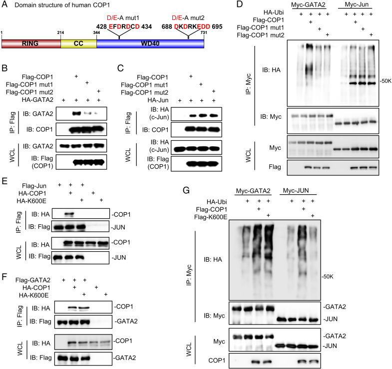
COP1 uses a typical mechanism to bind GATA2. (A) Domain structure of human COP1 and the two identified D/E-enriched motifs critical for COP1 binding to GATA2; CC, coiled coils; D/E-A, Mutations of EFDRDCD to AFARACA or DKDRKEDD to AKARKAAA; WD40 domain, a protein domain contains tandem copies of tryptophan–aspartate (WD) repeats of approximately 40 amino acids. (B) HEK293T cells were transfected with HA-tagged GATA2 (HA-GATA2) and Flag-tagged wild-type COP1 (Flag-COP1) versus two COP1 mutants (mut1 and mut2) with D/E-to-A mutations in the two D/E-enriched motifs as illustrated in A. After 48 h, cells were treated with 10 μM MG132 for 6 h before preparing the cell lysate for IP using anti-Flag affinity gel and IB analysis. (C) HEK293T cells were transfected with HA-tagged c-Jun (HA-Jun), Flag-COP1, Flag-COP1mut1, or Flag-COP1mut2. After 48 h, the cells were treated with 10 μM MG132 for 6 h before preparing the cell lysate for IP using anti-Flag affinity gel and IB analysis. (D) Ubiquitination assay. HA-Ubi and Myc-tagged GATA2 (Myc-GATA2) or c-Jun (Myc-Jun), along with Flag-COP1, Flag-COP1mut1, or Flag-COP1mut2 were co-transfected in HEK293T cells. After 48 h, the cells were treated with 10 μM MG132 for 6 h before preparing the cell lysate for IP using anti-Myc antibody and IB analysis. (E, F) HEK293T cells were transfected with HA-tagged COP1 (HA-COP1) or COP1K600E mutant (HA-K600E) together with (E) Flag-tagged c-Jun (Flag-Jun) versus control or (F) Flag-tagged GATA2 (Flag-GATA2) versus control. After 48 h, the cells were treated with 10 μM MG132 for 6 h before preparing the cell lysate for IP using anti-Flag affinity gel and IB analysis. (G) Ubiquitination assay. HA-Ubi and Myc-tagged GATA2 (Myc-GATA2) or c-Jun (Myc-Jun), along with Flag-tagged COP1 (Flag-COP1) or COP1K600E mutant (Flag-K600E) were co-transfected in HEK293T cells. After 48 h, the cells were treated with 10 μM MG132 for 6 h before preparing the cell lysate for IP using anti-Myc antibody and IB analysis. Data in B–G are shown as Western blots and are representative of at least three independent experiments; WCL, whole cell lysate.

