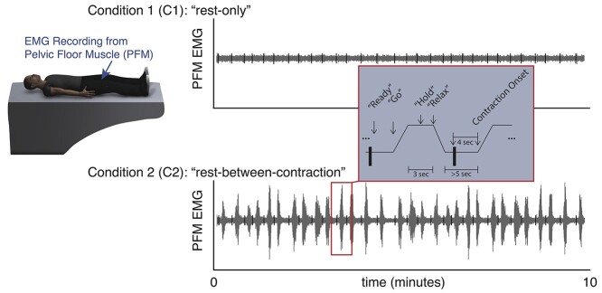
Figure 1.
Electromyographic (EMG) muscle activity to quantify the ability to relax the pelvic floor muscles (PFM). EMG recordings were made from the PFM. During condition 1 (C1) (“rest-only”), participants were instructed to rest quietly without going to sleep. During condition 2 (C2) (“rest-between-contraction”), participants repeatedly contracted and relaxed their PFM when cued by verbal instructions read by a computerized voice. Contraction onset times in the rest-between-contraction condition were calculated automatically from the EMG signal, and a series of 80-millisecond segments (black vertical bars) were examined 4 seconds prior to contraction onset during a period of time in which the participants had been instructed to relax. For consistency, the same segment timing was used to analyze both the rest-only condition and the rest-between-contraction condition.

