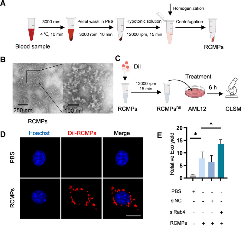
Fig. 3.
RCMP treatment boosts exosome yield. A Schematic illustration of RCMP preparation. B Representative TEM images of the prepared RCMP. C Schematic diagram of endocytosis of RCMPs into AML12 cells. D Fluorescence microscopy images of DiI-labeled RCMP (red) uptaken by AML12 cells. The cell nucleic was stained with Hoechst (blue). Scale bar = 5 μm. E RCMP treatment boost exosome yield. AML12 cells were treated with control or RCMPs or RCMPs together with siRNA and the exosomes produced were analyzed by NTA. All data are expressed as mean SEM of triplicate experiments. *p < 0.05

