Fig. 4.
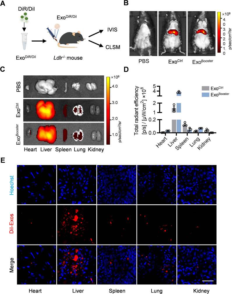
Biodistribution of ExoBooster in vivo. A Schematic illustration of the experimental procedure. B Representative IVIS images showing the distribution of the exosomes in vivo. Mice were injected with PBS or 100 μl DiR-labeled exosomes via tail vein. IVIS imaging was performed 4 h after injection. C Representative IVIS images of the DiR-labeled exosomes in different organs, including the heart, liver spleen, lung and spleen. D Quantification of the DiR signal intensity in Fig. 3C. n = 3. E Representative fluorescence microscopic images showing the distribution of exosomes in the tissue sections. Mice were injected with PBS or 100 μl DiI-labeled exosomes and different organs were harvested for tissue sectioning. The nuclei were counterstained with Hoechst. Scale bar = 50 μm

