Fig. 6.
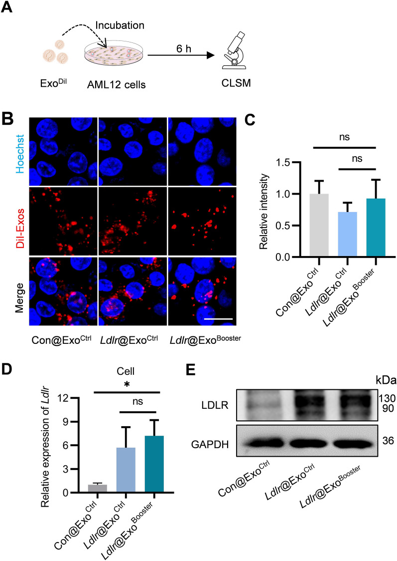
Ldlr@ExoBooster efficiently delivers therapeutic Ldlr mRNA into target cells. A Schematic diagram of the experiment. B Fluorescence images demonstrating the endocytosis of exosomes by the recipient cells. The distribution of DiI-labeled exosomes in AML12 cells were imaged by confocal microscopy, with the nuclei counterstained by Hoechst. Scale bar = 10 μm. C Fluorescence intensity of DiI signal corresponding to panel B. D qPCR analysis of Ldlr mRNA level in recipient cells. Data are expressed as mean SEM of three independent experiments. *p < 0.05 by one-way ANOVA. E Western blot analysis of LDLR expression at protein level in AML12 cells. Images are representative of three independent experiments

