Fig. 7.
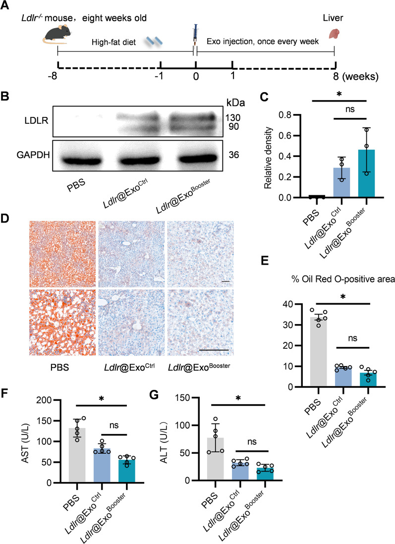
Ldlr@ExoBooster alleviates liver damage in Ldlr−/− mice. A Schematic illustration of the experimental procedure. Ldlr−/− mice were fed with high fat diet for 8 weeks, followed by PBS or exosome treatment once a week for 8 weeks. At the end of the experiments, mice were sacrificed and the liver tissues were harvested for systemic analysis. B Western blot analysis of LDLR protein expression in livers from mice treated as indicated. Data shown are representative of three independent experiments. C Quantification of western blot bands by densitometry. *P < 0.05 by one-way ANOVA. D Representative images of Oil Red O staining in liver slices from mice with indicated treatments. Scale bars = 50 μm. E Percentage of Oil Red O positive area in liver sections. Data are expressed as mean SEM. F, G Serum AST (F) and ALT (G) levels in the mice treated as indicated. *p < 0.05 by one-way ANOVA. n = 5. AST, aspartate aminotransferase; ALT, Alanine Aminotransferase

