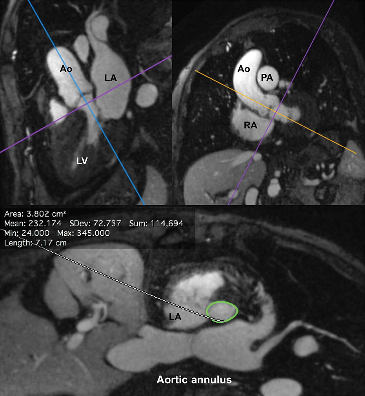
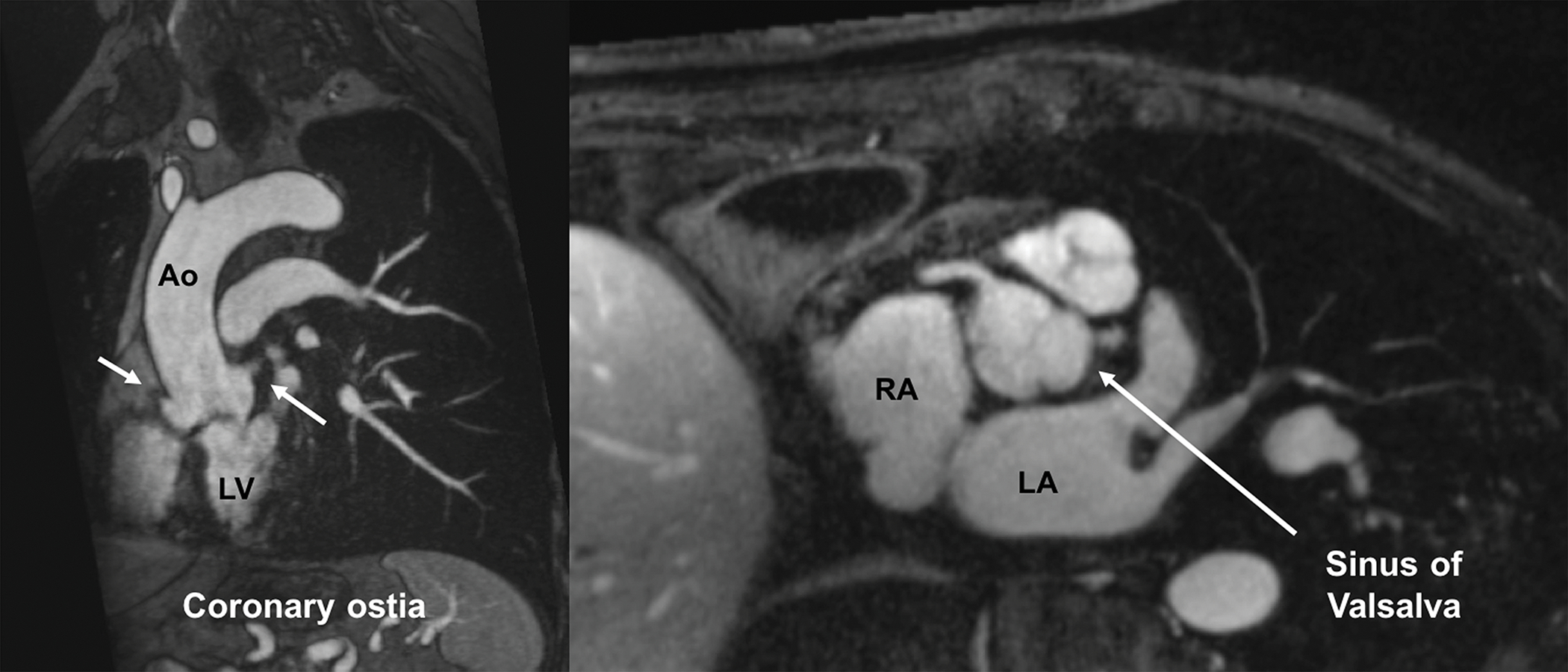
Figure 3.


FE-MRI for annular measurements and valve sizing. Multiplanar reformatted FE-MRA images are displayed and can be manipulated using a (a) double oblique technique to estimate transcatheter valve size and other vascular access sites. (b) Distance of the coronary arteries to the annular plane can also be assessed. White arrows show both right and left coronary ostia.
