
Figure 3.
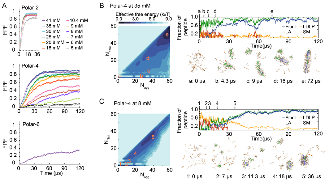
(A) Averaged-time course of the fraction of peptides in fibrils (FPF) for different concentrations of the polar-2, polar-4, and polar-6 model system, respectively. 2D PMF as a function of the aggregate size (Nagg) and corresponding number of peptides forming fibrils (Nfibril) for polar-4 at 35 mM and 8 mM are shown in the left of (B) and (C). Fraction of peptides in different components – i.e., low-density liquid phase (LDLP), spherical micelle (SM), large aggregates (LA) with Nagg > 9 in which the spherical micelles were excluded but nucleated fibril structures were included, and fibrils – as a function of simulation time are shown on the top right, and representative snapshots at different time points in the right bottom at 35 mM (B) and 8 mM (C). (Nagg, Nfibril) of each snapshot is marked as orange in the 2D PMF.