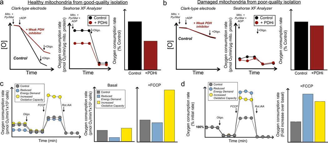
Figure 3 -. Importance of analyzing raw, quantitative rates and potential pitfalls of scaling respiration data.
(a) A hypothetical example is given where a weak inhibitor of pyruvate dehydrogenase (PDHi) reduces the ADP-stimulated respiration rate relative to vehicle controls in isolated mitochondria offered pyruvate with malate, as shown in both a (left) Clark-type electrode and (middle) Seahorse XF Analyzer. (right) Data normalized to 100% of the vehicle control. Mito., isolated mitochondria; Oligo., oligomycin; PDHi, pyruvate dehydrogenase inhibitor. (b) The same experiment conducted with mitochondria from a suboptimal isolation yields different results. With low rates from damage during the isolation, the effect of PDHi is no longer rate-controlling. Presenting percentage-based data relative to vehicle control shows little difference between groups and the confounding effects of the poor isolation cannot be identified. Abbreviations as before. (c) A hypothetical example is presented of two potential cellular phenotypes that may arise from genetic transformation or pharmacologic intervention. (left) A reduction in ATP demand (blue) lowers the basal rate of respiration but leaves FCCP-stimulated respiration largely unchanged. Additionally, an increase in oxidative capacity (yellow), as can happen with enhanced mitochondrial biogenesis, has little effect on basal respiration but a dramatic effect on the maximal respiration. (right) Calculating the quantitative parameters easily identifies the respective changes. Oligo., oligomycin; Rot/AA, rotenone with antimycin A. (d) Scaling the rates to the initial rate of respiration masks the precise changes, as both present with roughly equal fold-changes in uncoupler-stimulated respiration. Abbreviations as before.

