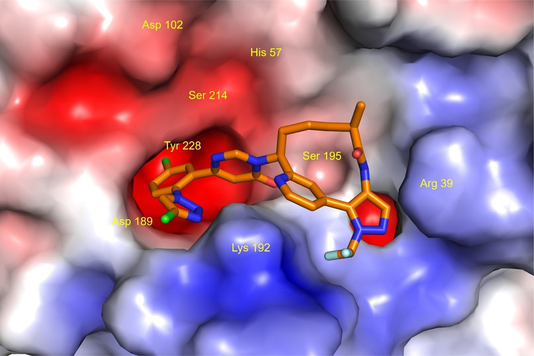
Fig. 2. X-ray crystal structure of milvexian bound to factor XIa (Protein Data Bank ID: 7MBO).

Milvexian is represented as a stick model (atom colors: C, orange; O, red; N, blue; Cl, green; F, light blue; H, not shown). The molecular surface of Factor XIa is displayed colored by electrostatic potential (blue: positive; red: negative; white: neutral). Amino acid labels are displayed for the catalytic triad residues (His57, Asp102, Ser195) and selected residues (Arg39, Asp189, Lys192, Tyr228) that are within a distance of 3.5 angstroms from milvexian. This illustration was created using the PyMOL Molecular Graphics System, version 1.7.6.0, Schrödinger, LLC, New York.
