
Figure 3.
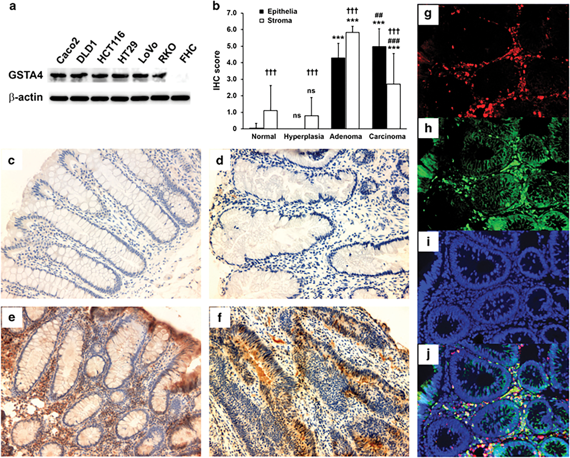
GSTA4 is expressed in human colon adenomas and carcinomas. (a) Western blots show increased expression of GSTA4 in colon cancer cell lines compared to a fetal human colonic epithelial cell line (FHC). (b) Immunohistochemical staining scores for GSTA4 in human colon tissues (NS, not significant, ***P < 0.001 compared to TANC biopsies; ##P < 0.01 and ###P < 0.001 compared to tubular adenomas; †††P < 0.001 compared to epithelia). (c-f) Representative immunohistochemical staining of colon biopsies for GSTA4 in TANCs (c), hyperplastic polyps (d), tubular adenomas (e), and invasive carcinomas (f); intense staining is evident for GSTA4 in tubular adenomas and invasive carcinomas compared to normal tissue and hyperplastic polyps. (g-j) Immunofluorescent staining for F4/80 (g, red) and GSTA4 (h, green) shows co-localization of GSTA4 and macrophages (j, yellow) in a tubular adenoma. Nuclei are counterstained with DAPI (i and j, blue).