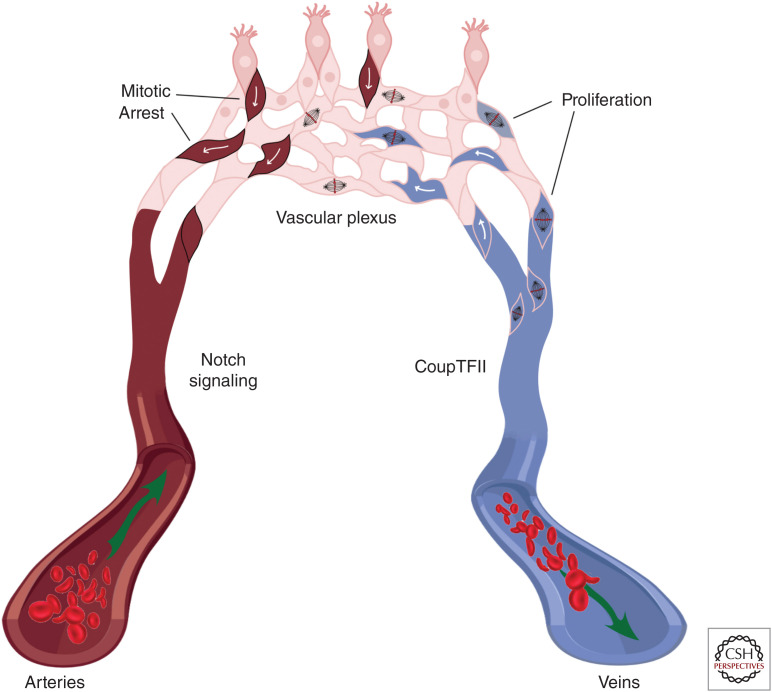
Figure 2.
Arterial/venous differentiation from the vascular plexus. Arterial endothelial cells (ECs) (left) express Notch proteins, while venous ECs (right) have low Notch expression and high CoupTFII expression. CoupTFII drives endothelial proliferation in venous endothelium, extending the vein and contributing cells to the growing vascular plexus (top). Stalk cells in the vascular plexus experience high levels of Notch signaling, which causes cell-cycle arrest. High Notch signaling and cell-cycle arrest leads the cell to assume arterial fate, migrate against the direction of blood flow, and incorporate into the artery.

