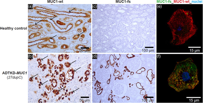
FIGURE 3.

Immunostaining of previously obtained kidney biopsies or urinary cells can be used to identify MUC1 frameshift (MUC1‐fs) protein in cases where the mutation cannot be found genetically. (a) Horseradish peroxidase staining of kidney tissue from a healthy control with antibody to the normal mucin‐1 (MUC1‐wt). One can see apical staining of the MUC1 protein (black arrow), consistent with apical expression of MUC1‐wt. (b) Similar immunostaining for ADTKD‐MUC1: immunostaining shows a clumping of deposits within the cell and absence of normal apical staining (black arrow). (c) Staining with an antibody for the MUC1‐fs protein in the healthy control. No staining is found. In contrast, in (d) immunostaining with the MUC1‐fs is positive in ADTKD‐MUC1. (e, f) Staining of urinary cells for the MUC1‐wt protein (red) and the MUC1‐fs protein (green). In the healthy control, there is only positive staining for the MUC1‐wt (all staining in red). In (f) one sees the MUC1‐fs protein deposited intracellularly. The MUC1‐wt is localized partially at the cytosol and partially on plasma membrane in the urinary cells
