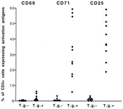
FIG. 2.
Percentages of CD3+ lymphocytes expressing CD69, CD71, and CD25 above control levels after in vitro stimulation with soluble T. gondii antigen in whole blood from 10 T. gondii-positive (T.g.+) and 10 T. gondii-negative (T.g.−) individuals. Stimulation and staining were performed as described in Materials and Methods. Each symbol represents one individual. Statistical analysis (Mann-Whitney U test) showed significantly higher percentages of CD3+ CD25+ and CD3+ CD71+ cells than of CD3+ CD69+ cells in T.g.+ individuals (P < 0.0001 in each case).

