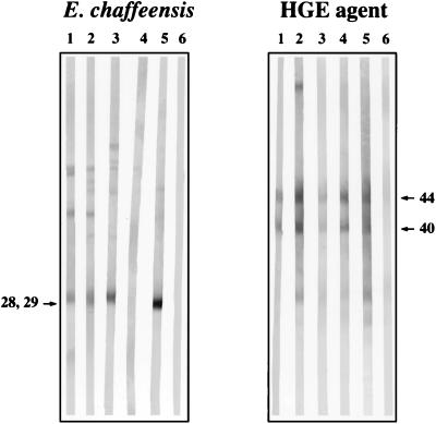
FIG. 1.
Immunoblotting to differentiate E. chaffeensis and HGE agent antibodies in deer sera. (Left panel) Lane 1, Maryland sample 8 (E. chaffeensis IFA positive only); lane 2, Maryland sample 10 (E. chaffeensis and HGE agent IFA positive); lane 3, Wisconsin sample C8 (E. chaffeensis and HGE agent IFA positive); lane 4, Wisconsin sample SP10 (E. chaffeensis and HGE agent IFA-negative control); lane 5, mouse anti-E. chaffeensis monoclonal antibody 1A9; lane 6, normal mouse serum. (Right panel) Lane 1, Wisconsin sample C9 (HGE agent IFA positive only); lane 2, Wisconsin sample C43 (HGE agent IFA positive only); lane 3, Wisconsin sample C8 (E. chaffeensis and HGE agent IFA positive); lane 4, Maryland sample 10 (E. chaffeensis and HGE agent IFA positive); lane 5, Wisconsin sample SP15 (HGE agent IFA-positive control); lane 6, Wisconsin sample SP10 (E. chaffeensis and HGE agent IFA-negative control). Numbers beside the gels are molecular sizes of the diagnostically significant 28- to 29-kDa antigen of E. chaffeensis and 44-kDa antigen of the HGE agent.

