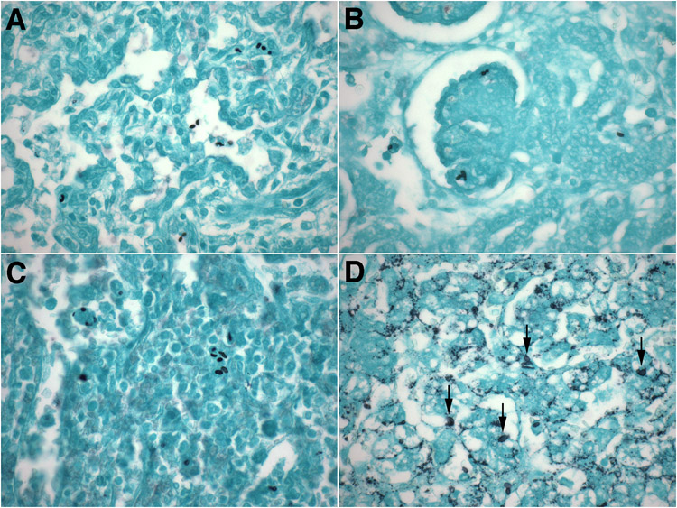
Figure 1. Anatomic distribution of yeasts in invasive neonatal non-albicans candidiasis.
A-D. Representative micrographs of lungs (A), kidneys (B), spleen (C) and liver (D) of extremely preterm neonates with invasive non-albicans candidiasis showing the ubiquitous presence of Candida yeast forms (2-5 μm ovals) in these organs. While readily visible by Gomori-methenamine silver staining in most organs (A-C), the Candida yeasts were obscured by intrinsic lipochrome pigment in liver (D, arrows).
(A-D: Gomori-methenamine silver, original magnification: x600).

