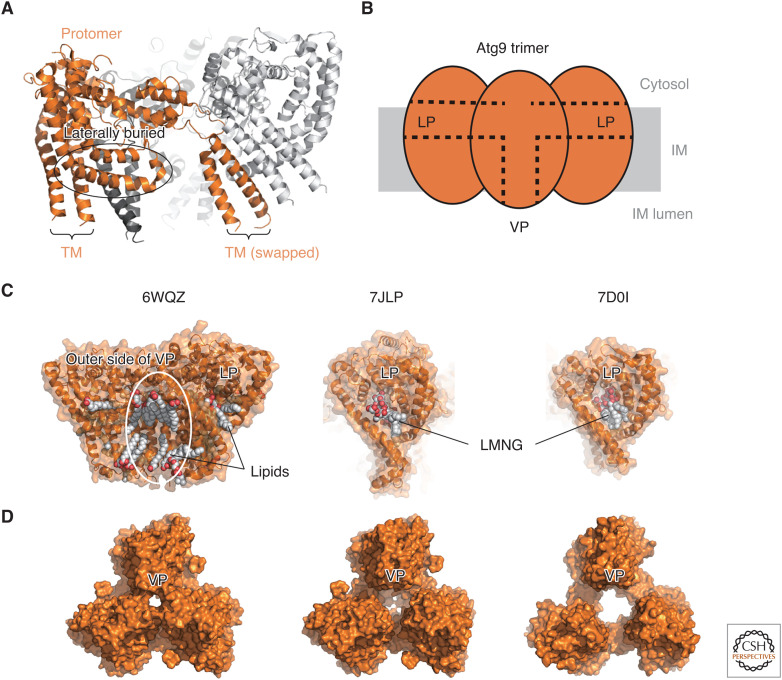
Figure 3.
Structural basis of Atg9-mediated lipid scrambling. (A) Ribbon model of fission yeast Atg9 (PDB 7D0I). One protomer is colored orange, whereas the other two protomers within a trimer are colored light and dark gray, respectively. (TM) Transmembrane helices. (B) Schematic drawing of Atg9 trimer. (C) Lipids and detergents bound to the lateral pore (LP) and vertical pore (VP) of Atg9/ATG9A. (Left) Lipids bound to the outer side of VP and LP of human ATG9A (PDB 6WQZ). (Middle) LMNG bound to the LP of human ATG9A (PDB 7JLP). (Right) LMNG bound to the LP of fission yeast Atg9. (D) Various-sized VPs observed at the center of the Atg9/ATG9A trimers.

