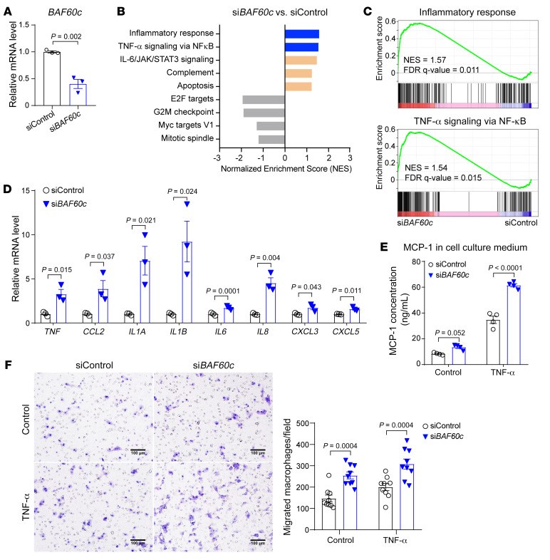
Figure 5. BAF60c depletion enhances VSMC inflammatory response.
(A–C) HASMCs were transfected with siControl or siBAF60c (30 nM). After 24 hours, cells were serum starved in Opti-MEM for 24 hours, followed by total RNA extraction for qPCR (A) and for RNA-Seq (B and C). (B) GSEA was performed comparing siBAF60c with siControl. (C) Positive enrichment of the inflammatory response (left) and TNF-α signaling via NF-κB (right) in GSEA plots (siBAF60c vs. siControl). (D) qPCR to validate the expression of inflammation-related genes in 3 independent sets of samples treated as in A. (E) MCP-1 concentration was measured by ELISA in cell culture media. HASMCs were transfected with siControl or siBAF60c. After 48 hours, cells were cultured in opti-MEM with or without TNF-α (20 ng/mL) for 24 hours. Data are from 4 independent experiments. (F) Representative images (magnified field, left) and quantitative analysis (right) of BMDMs (isolated from WT mice) in the Transwell migration assay cocultured with HASMCs transfected with siControl or siBAF60c in the presence or absence of TNF-α (20 ng/mL) (n = 9 images/group). Scale bars: 100 μm. Data are represented as mean ± SEM. Student’s t test (A and D); 2-way ANOVA followed by Holm-Šidák post hoc analysis (E and F).

