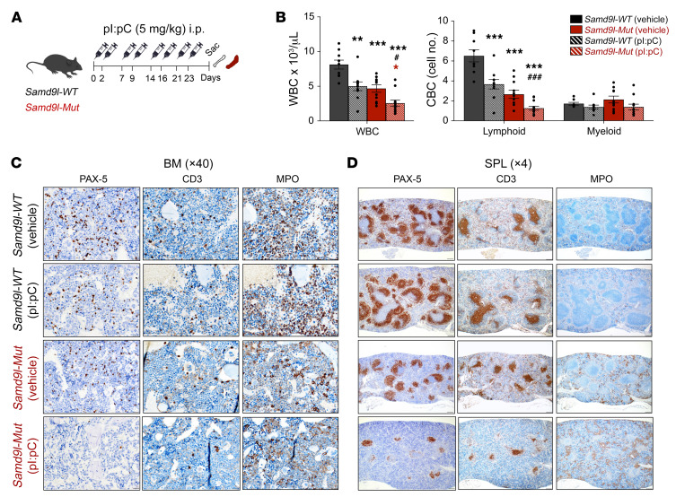
Figure 6. Inflammation worsens cytopenias in Samd9l-Mut mouse.
(A) Illustration for treatment of Samd9l-WT and Samd9l-Mut mice with pI:pC or vehicle. (B) CBC analysis showing WBC, lymphoid cells, and monocytes/neutrophils/basophils (myeloid) for the treated mice (n = 8). (C and D) BM (C) and spleen (D) were stained with anti–PAX-5, anti-CD3, and anti-MPO to assess B, T, and myeloid cells, respectively (n = 2 per group). For panel B, the Kruskal-Wallis test was used to perform an initial comparison across all groups, and followed by pairwise comparisons with Wilcoxon’s rank-sum test. *P < 0.05, **P < 0.01, ***P < 0.001 compared with vehicle-treated groups. #P < 0.05, ###P < 0.001 compared with pI:pC-treated groups. Color indicates the comparison group. Error bars indicate the SEM for biological replicates. Black, Samd9l-WT, red, Samd9l-Mut; stripes, pI:pC; solid, vehicle.

