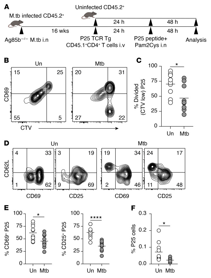
Figure 6. Chronic pulmonary M. tuberculosis infection impairs naive CD4+ T cell response to an unrelated antigen in mLN.
(A) Experimental design. B6 mice were infected with Ag85b–/– M. tuberculosis intranasally (i.n.) or remained uninfected. At 16 weeks p.i., infected and uninfected mice received i.v. CTV-labeled splenocytes from Ag85b-specific P25.CD45.1+.Rag1–/– TCR-Tg mice (~5 × 10⁵ CD4+ T cells per recipient). Twenty-four hours later, both groups of mice were inoculated with 10 μg Ag85b240–254 peptide together with Pam2Cys adjuvant i.n. The mLNs were collected 48 hours after inoculation. (B and C) Flow cytometry plots showing the expression of CD69 on CTV-labeled P25 cells (B) and summary graph of the proportion of divided P25 cells (CTV low) (C) in mLN of uninfected (Un) and infected (Mtb) mice. (D) Representative flow cytometry plots showing the expression of CD62L, CD69, and CD25 on P25 cells. (E) Summary graphs showing the proportion of CD69+ and CD25+ P25 cells in mLN of uninfected and M. tuberculosis–infected mice. (F) The proportion of P25 cells in total CD4+ T cells in infected and uninfected mice. Data shown are from 2 independent experiments (n = 10 mice per group in total). Statistical differences between groups were determined using Student’s t test. *P < 0.05, ****P < 0.0001.

