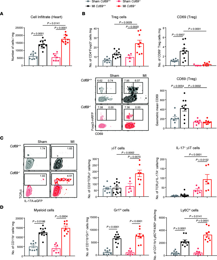
Figure 4. Myocardial accumulation of CD69+ Tregs and Il-17+ γδT cells after LAD ligation.
(A) Leukocyte cell numbers per milligram of heart tissue in the myocardium 2 days after infarction. (B) Quantification of the number of Tregs (CD4+Foxp3+) and CD4+Foxp3+CD69+ cells per milligram of heart tissue and CD69 mean fluorescence expression on Tregs in the heart. Representative density plots showing gating on CD45+CD11b–CD4+ cells. (C) Representative density plots gated on CD45+CD11b–CD3+ cells and numbers of γδT cells and Il-17+ γδT cells per milligram of tissue. (D) Quantification of total cell numbers per milligram of CD11b+ myeloid cells, CD11b+Gr1hi cells, and CD11b+F4/80loLy6Chi cells in the heart. Heart cell–infiltrating populations were evaluated 2 days after infarction (n = 6–11 animals per group). Data are representative of 4 independent experiments and indicate the mean ± SEM. Statistical significance was analyzed by 1-way ANOVA with Tukey’s post hoc test. P values for significant differences are shown.

