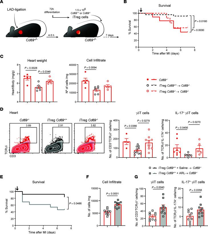
Figure 6. Adoptive transfer of CD69-sufficient Tregs into Cd69–/– mice reduces myocardial inflammation and improves survival after MI.
(A) Schematic workflow of the iTreg adoptive transfer after LAD ligation. (B) Survival after LAD ligation (n = 7–24). Black arrow depicts the time of iTreg inoculation (4–5 hours after infarction). Cd69–/– mice without cell transfer were used as controls. The P value was calculated by long-rank (Mantel-Cox) test. (C) Heart/body weight ratio and total leukocyte numbers per milligram of heart tissue 7 days after LAD ligation (n = 5–6). (D) Representative density plots (gated on CD45+CD11b– cells) and numbers of γδT cells and IL-17+ γδT cells per milligram of myocardial tissue. Data in C and D correspond to 1 representative independent experiment of 3 experiments. Data indicate the mean ± SEM and were analyzed by 1-way ANOVA with Tukey’s post hoc test. (E) Survival after LAD ligation (n = 12–13), Cd69+/+ iTreg transfer, and ARL 67156 or saline administration. Black arrow depicts the time of iTreg inoculation (4–5 hours after infarction). The P value was calculated by long-rank (Mantel-Cox) test. (F) Total leukocyte numbers per milligram of heart tissue 7 days after LAD ligation (n = 9). (G) Numbers of γδT cells and IL-17+ γδT cells per milligram of myocardial tissue (n = 9). Data in F and G were pooled from 4 independent experiments and indicate the mean ± SEM. Statistical significance was determined by unpaired Student’s t test.

