. 2022 Oct 31;12(48):31068–31082. doi: 10.1039/d2ra05127c

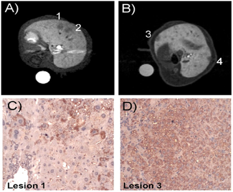
Fig. 4. Histological validation of MRI lesion detection. T1-weighted MR images (liver region), of HBV-Tg transgenic mice 20 min after the injection of Mn-Apo (A) and Gd–BOPTA (B), respectively. Lesions 1 and 2 were easily detectable only after Mn-Apo administration. However, lesions 3 and 4 were detectable only after Gd–BOPTA injection. (C and D) Expression pattern of SCARA5 in HBV-Tg. (C) Immunohistochemical staining of lesion 1 showing low Mn-Apo uptake. SCARA5 was observed only in some HCC cells. (D) Immunohistochemical staining of lesion 3 showing higher Mn-Apo uptake. SCARA5 was present in almost all cancer cells. Reproduced with permission from ref. 50. Copyright John Wiley & Sons, Ltd, 2012.

Fig. 4