. 2022 Oct 31;12(48):31068–31082. doi: 10.1039/d2ra05127c

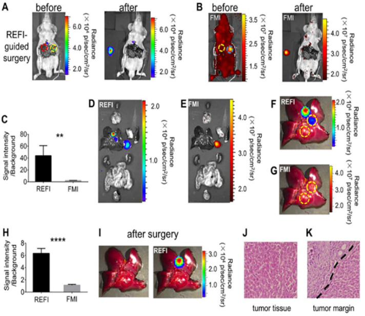
Fig. 5. REF image-guided cancer surgery of the orthotopic HCC tumor-bearing mice. (A) Representative REFI of the mouse after the laparotomy (left). After the REF image-guided cancer surgery, the mouse and the resected tumor were also imaged for REFI (right). (B) Representative fluorescent images of the mouse before and after the tumor resection. (C) Comparison of SNR of REFI and FMI. (D) and (E) REFI and FMI of all the organs and the orthotopic HCC. (F) and (G) Representative REF and FM image of the mouse liver, respectively. (H) Comparison of quantification of SNR of REFI and FMI of the mouse liver. (I) Photograph and REF image of the mouse liver after the tumor second resection, respectively. (J) and (K) H&E of the orthotopic HCC and surgical margin, respectively. Reproduced with permission from ref. 61. Copyright Elsevier Inc, 2017.

Fig. 5