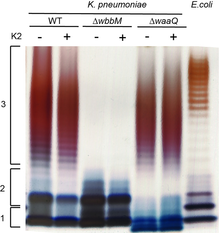
Fig 10. K2 treatment does not visibly affect LPS structure.
Bacteria were grown overnight in the presence or absence of K2. LPS from K. pneumoniae WT and mutant strains (ΔwbbM, ΔwaaQ) were then purified and analyzed by SDS-PAGE electrophoresis and silver staining. LPS extracted from E. coli (serotype O111:B4) was used for comparison. The LPS of WT K. pneumoniae showed the three main forms of the LPS: lipid A (1), lipid A+ oligosaccharide core (2), lipid A + core + O-antigen [27]. As expected, the LPS from ΔwbbM bacteria lacked the O-antigen, and ΔwaaQ displayed a smaller oligosaccharide core that migrated further in the gel. No visible alteration of LPS structure was observed in K2-treated bacteria.

