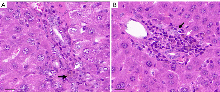
Figure 2.
Histopathologic evaluation of rat livers. (A) The bile duct cells are disorderly arranged (black arrow) and infiltrated with inflammatory cells (white arrow). The pathological grade was 3. (B) The bile duct cells are damaged (black arrow), with cholestasis and a large number of infiltrated inflammatory cells in the periphery (white arrow). The pathological grade was 4. H&E staining magnification: ×400, scale bar 20 µm. H&E, hematoxylin and eosin.

