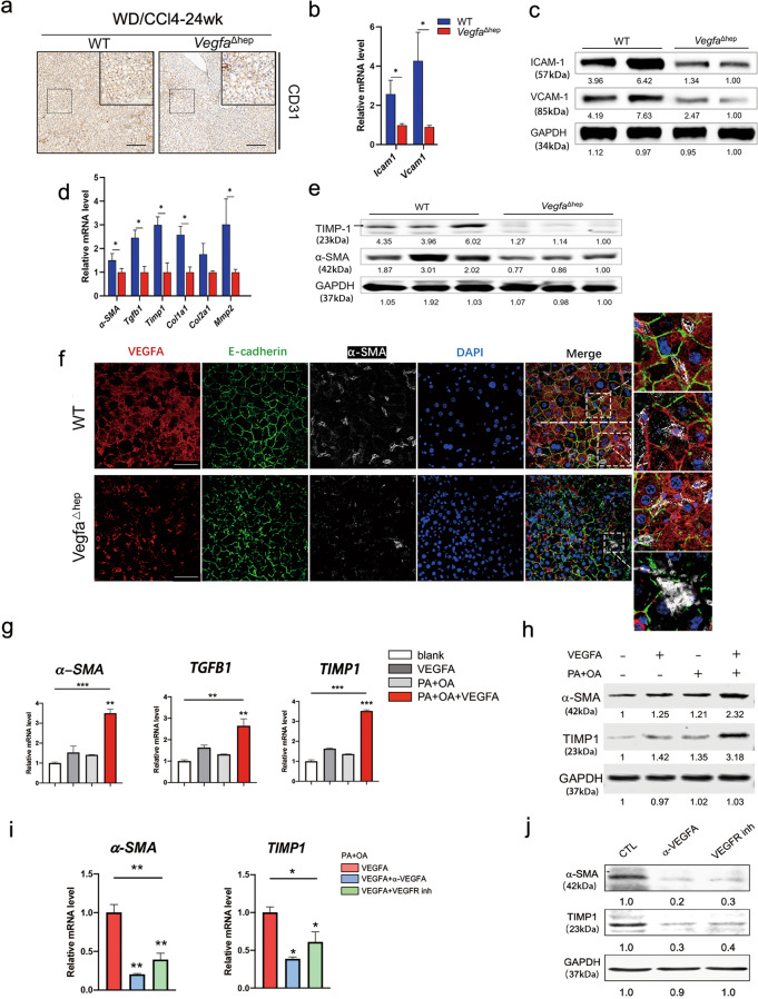
Fig. 4. Hepatocyte-derived VEGFA promoted endothelial dysfunction and activated HSCs during NAFLD-HCC transition.
a Representative immunohistochemical staining of CD31 in WD/CCl4-WT and -VegfaΔhep livers at 24 weeks. Scale bars, 100 µm. b, c RNA and protein expression of endothelial dysfunction markers in WD/CCl4-WT and -VegfaΔhep mice were measured by real-time PCR (b) and Western blot (c). (n = 8). Data are represented as mean ± SD. d, e RNA (d) and protein expression (e) of activated HSCs markers in WD/CCl4-WT and -VegfaΔhep mice were measured. (n = 8). f Immunofluorescence analyses of VEGFA expression and HSCs activation in WD/CCl4-WT and -VegfaΔhep mice at 24 weeks. Activated HSCs were immunostained by α-SMA. Scale bars, 25 µm. g, h Relative RNA (g) and protein (h) levels of activated HSCs-related markers in LX2 treated with VEGFA (50 nM), PA (0.5 mM) + OA (1 mM), VEGFA + PA + OA, versus with Vehicle control for 24 h, respectively. i, j Relative RNA (i) and protein (j) levels of activated HSCs-related markers in LX2, which was pretreated with VEGFA + PA + OA, then treated with α-VEGFA (2 μg/mL) or VEGFR inhibitor Axitinib (0.3 nM, VEGFR inh), respectively. One-way ANOVA followed by Tukey’s test was used for multiple comparisons. *P < 0.05, **P < 0.01, ***P < 0.001.

