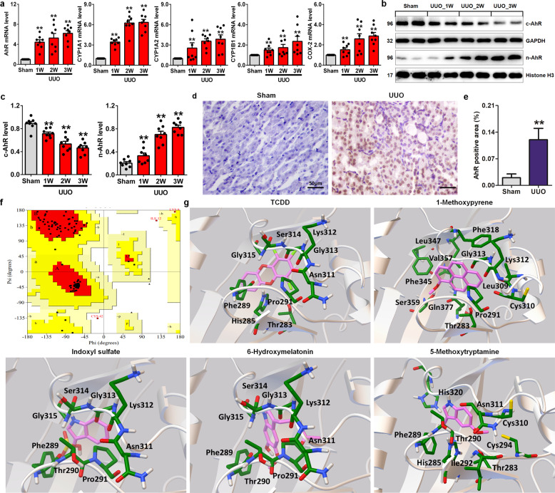
Fig. 3. AhR is activated by endogenous aryl-containing metabolites in the progressive TIF of UUO mice.
a Time course of the mRNA expression of AhR and its target genes, including CYP1A1, CYP1A2, CYP1B1 and COX-2, in the obstructed kidney tissues of UUO mice at Weeks 1, 2 and 3. b Protein expression of AhR in the nucleus and cytoplasm of the obstructed kidneys of UUO mice at Weeks 1, 2 and 3. c Quantitative analysis of AhR in the nucleus and cytoplasm of the obstructed kidneys of UUO mice at Weeks 1, 2 and 3. d Immunohistochemical analysis with anti-AhR in the obstructed kidneys of UUO mice. e Quantitative results of AhR expression in the obstructed kidneys of UUO mice. f Ramachandran plot of the modelled mouse AhR-LBD. g The predicted binding mode of the four metabolites and TCDD docked into AhR-LBD (grey ribbon form). Stick structures represent the amino acid residues of AhR-LBD. *P < 0.05, **P < 0.01 vs the sham group.

