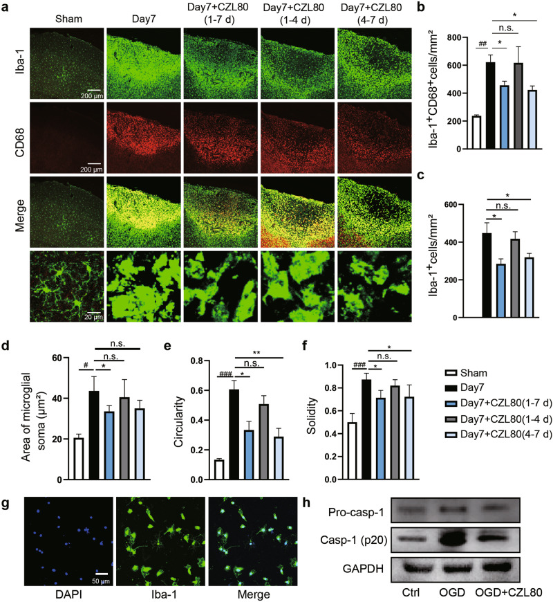
Fig. 7. CZL80 inhibits the activation of microglia.
a Immunofluorescent staining of Iba-1-positive (green) and CD68-positive (red) microglia in and around the ischemic core area. Representative images (a) and quantitative analysis (b, c) showing the effect of CZL80 administration in different time periods on the immunostaining of Iba-1 and CD68 after PT. Statistical significance was determined using one-way ANOVA with Tukey’s post hoc test, n.s. (not significant vs. Day7). ##P < 0.01 vs. Sham, *P < 0.05 vs. Day 7. d–f Quantitative analysis of the effects of CZL80 on soma area of microglia (d, #P < 0.05 vs. Sham, *P < 0.05 vs. Day 7), circularity (e, ###P < 0.001 vs. Sham, *P < 0.05, **P < 0.01 vs. Day 7) and solidity (f, ###P < 0.001 vs. Sham, *P < 0.05 vs. Day 7). g The primary cultured microglial cells were identified by staining with the microglia marker Iba-1. h CZL80 suppressed the expression of Caspase-1 in primary cultured microglia after OGD/R, as measured by western blot. All of the data are shown as mean ± SEM. These experiments were repeated at least for 3 times independently.

