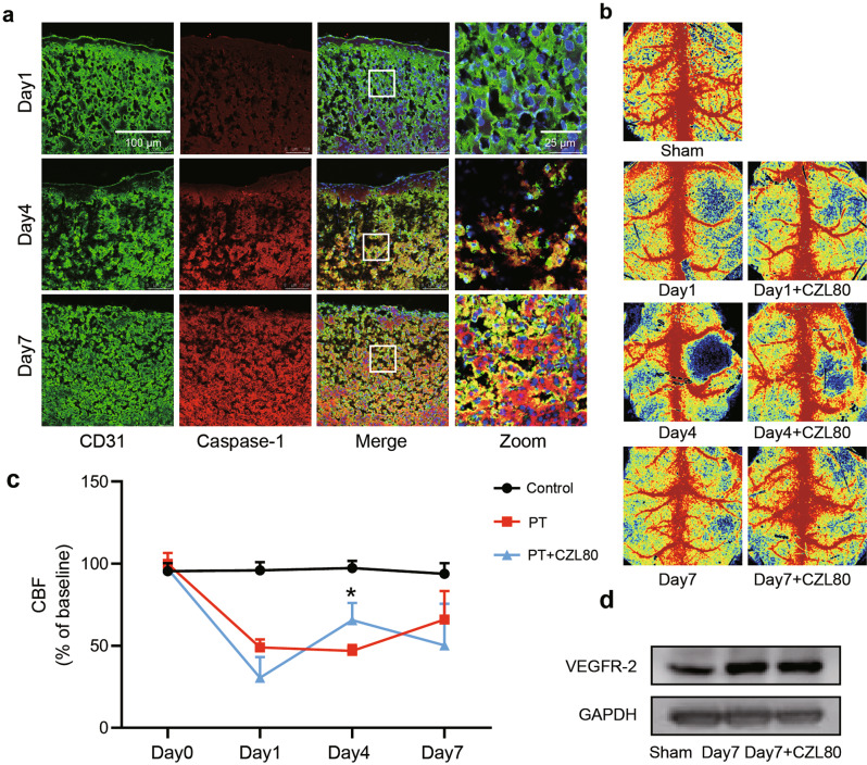
Fig. 8. Angiogenesis and improved blood flow have limited contribution to the pharmacological effects of CZL80.
a Representative images showing the immunofluorescent staining of Caspase-1 with endothelial cells on Day1, 4, 7 after photothrombosis. CD31 was taken as markers of endothelial cells (CD31). b Representative cerebral blood flow images from indicated groups. c Statistical data showing changes in CBF for the Sham group, PT group and PT + CZL80 group on Day0, Day1, Day4 and Day7. Results were represented as means ± SEM (n = 5–6 animals per group). Statistical significance was determined using one-way ANOVA with Tukey’s post hoc test, *P < 0.05 vs. PT group. d Representative immunoblots showing VEGFR-2 in the peri-infarct cortex from indicated groups. These experiments were repeated at least for 3 times independently.

