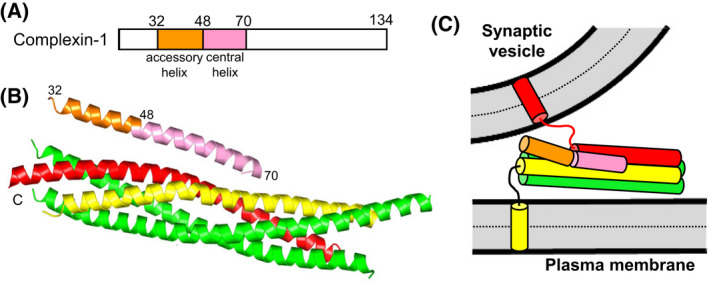
Fig. 3.

Complexin‐1 structure and function. (A) Domain diagram of complexin‐1. Selected residue numbers are indicated above the diagram. (B) Ribbon diagram of the crystal structure of Cpx1(26–83) (accessory helix in orange, central helix in pink) bound to the SNARE complex, with synaptobrevin in red, syntaxin‐1 in yellow and SNAP‐25 in green [43] (PDB accession number 1KIL). Selected residue numbers are indicated. C denotes the C‐termini of the SNAREs. (C) Model showing how the accessory helix of complexin‐1 bound to a partially assembled trans‐SNARE complex is expected to have steric clashes with the vesicle that would hinder final C‐terminal zippering of the SNARE four‐helix bundle [84].
