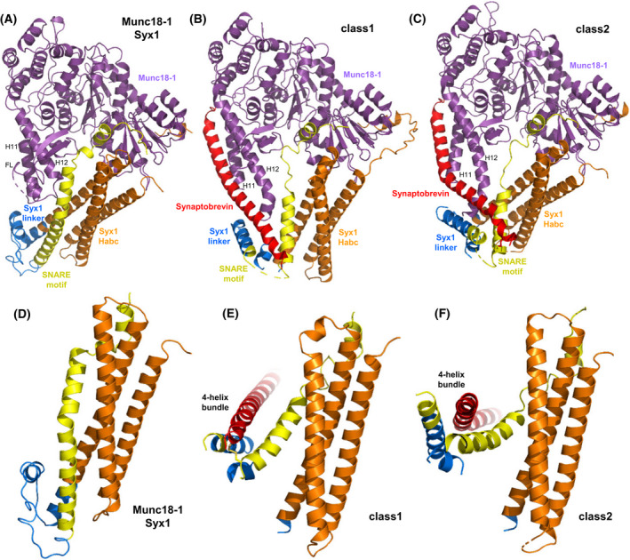
Fig. 7.

Function of Munc18‐1 in organizing SNARE complex assembly. (A–C) Ribbon diagrams of the crystal structure of the Munc18‐1‐closed syntaxin‐1 complex (A) [20] and the two cryo‐EM structures of the Munc18‐1‐syntaxin‐1‐synaptobrevin template complex (B, C) [33] (PDB accession numbers 3C98, 7UDC and 7UDB, respectively). Syntaxin‐1 is abbreviated Syx1 and the two cryo‐EM structures are denoted class1 and class2. Munc18‐1 is in purple, synaptobrevin in red and syntaxin‐1 in orange (N‐peptide and Habc domain, blue (linker region) and yellow (SNARE motif). The positions of helices 11 and 12 (H11 and H12) of Munc18‐1, and the furled loop that connects these helices and hinders synaptobrevin binding (labeled FL) are indicated. (D–F) Close‐up views of the region where the syntaxin‐1 SNARE motif contacts the Habc domain and the linker in the Munc18‐1‐closed syntaxin‐1 complex (D), class1 (E) and class2 (F). Munc18‐1 is not shown for simplicity. Note how the SNARE motif separates gradually from the Habc domain in the three structures from left to right and how the syntaxin‐1 linker forms a short four‐helix bundle with the syntaxin‐1 and synaptobrevin SNARE motifs in class1 and class2.
