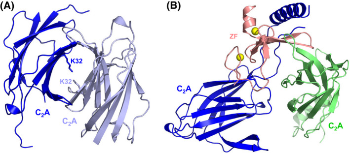
Fig. 9.

Munc13‐1‐RIM homodimer heterodimer switch. (A, B) Crystal structures of the Munc13‐1 C2A domain homodimer (A) and the Munc13‐1 C2A domain‐Rim ZF heterodimer (B) [59] (PDB accession numbers 2CJT and 2CJS, respectively). In (A), the K32 side chains in the homodimerization interface are labeled. In (B), the RIM ZF domain is in salmon and the two Munc13‐1 fragments containing the C2A domain observed in the crystals are in blue and light green. Zinc ions are shown as yellow spheres. Note that the interface of the blue fragment with RIM is the biologically relevant interface [183], whereas the interfaces formed by the light green fragment arise because of crystal contacts [59].
