Figure 3.
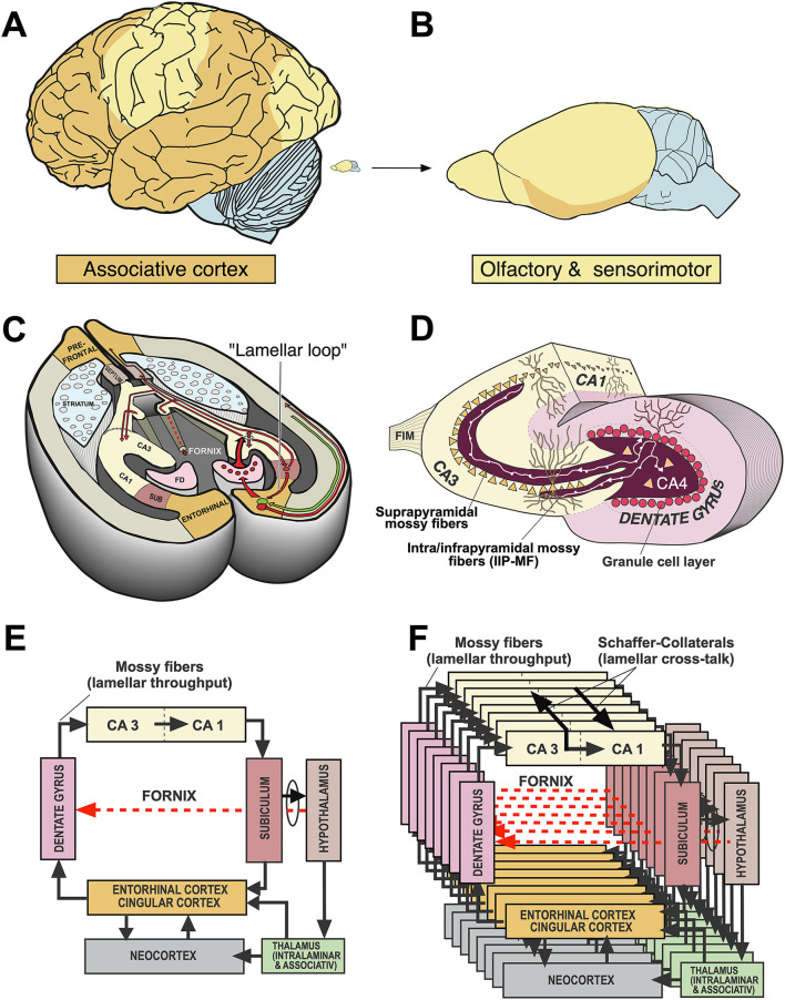
Comparative aspects of hippocampal circuitry. (A) In humans, a huge part of the neocortex is devoted to associative cortex, while cortical regions with specialized sensory or motor function are relatively restricted. (B) The rat neocortex includes mostly specialized sensorimotor areas but also olfactory input. Associative cortex is reduced and appears marginal. (C) The horizontal section through a mouse brain reveals that the associative cortices fold in to form a relatively large hippocampal formation that is topped at its end by the (late developing) dentate gyrus. The right half shows the typical feed-forward loop originating from the entorhinal cortex that transmits topographically ordered input (green arrow) from the remaining associative cortex as perforant path to the granule cells (red) in the dentate gyrus. From there, mossy fibers run along the pyramidal cells of hippocampal subregion CA3. The targeted neurons in CA3 give off axonal branches known as Schaffer collaterals that transmit the inputs to CA1 and eventually to the subiculum. The subiculum gives collaterals to the entorhinal cortex, closing the so-called lamellar loop, but is also sending fibers via the fornix that end in various subcortical structures, such as septum and hypothalamus. Note that the fornix contains also ascending fiber projections (see below). While the lamellar loop appears similarly organized in both species, the rodent hippocampus shows massive commissural projections largely lacking in humans in which commissures connect the entorhinal cortices. (D) 3D-scheme of the hippocampal mossy fiber projections. Axons of the granule cells run along the CA3 region contacting numerous pyramidal cells with so-called giant boutons. (E) Planar scheme of the lamellar (“trisynaptic”) loop starting at the entorhinal cortex, with the subiculum as the main output structure to both cortical and (via Fornix) to subcortical structures along the basal forebrain. The red arrow symbolizes the ascending projection from the hypothalamus to the hippocampus (for details see Figure 13B). (F) Stacked trisynaptic lamellae showing the lateral spread of the Schaffer collaterals, probably less in CA3 than in CA1. FD, fascia dentata (dentate gyrus); FIM, fimbria hippocampi; SUB, subiculum.

