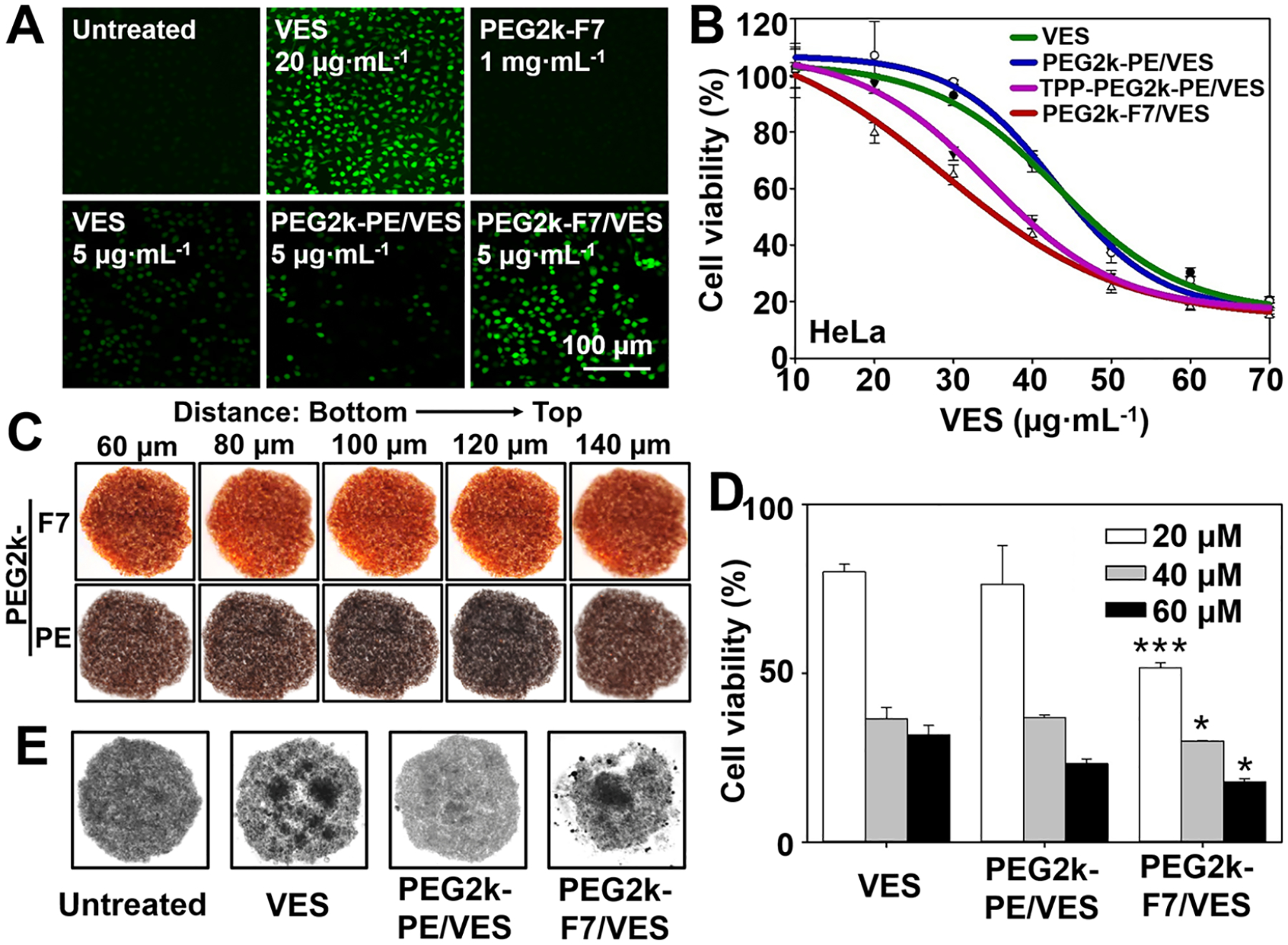
Figure 4.

In vitro drug delivery. (A) Cellular ROS levels. Cells were incubated with various formulations for 20 h, and then incubated with the H2DCFDA (10 μM) at 37 °C for 0.5 h. The generated green fluorescence, an indicator of the ROS, was determined by fluorescence microscopy. (B) Cytotoxicity of the VES-loaded micelles determined by the CellTiter-Blue Cell Viability assay. Incubation time: 48 h. (C) Penetration of the Rh-PE-labeled micelles in the cell spheroids. Incubation time: 1 h. (D) Cytotoxicity and (E) spheroid morphology upon treatments in the cell spheroids. Incubation time: 72 h. Cell line: HeLa cells. Data were expressed as the mean ± SD, *p < 0.05; ***p < 0.001, when compared to the PEG2k-PE/VES group.
