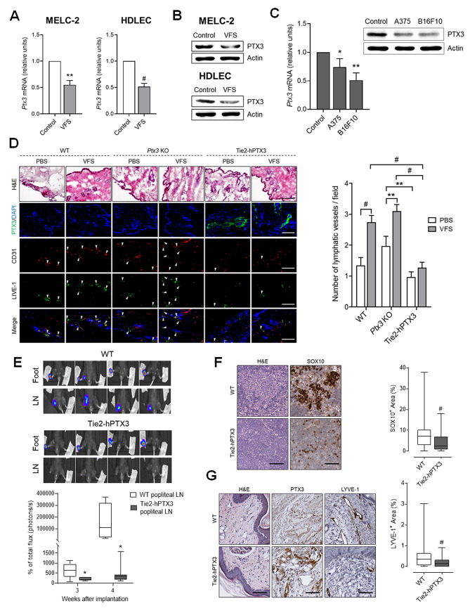
Fig. 2.
PTX3 impairs the lymphangiogenesis and lymphatic dissemination. qPCR of PTX3 mRNA (A) and Western blot analysis (B) of MELC-2 and HDLEC treated with VFS or not (control). C) qPCR (left panel) and Western blot analysis (right panel) for PTX3 expression in HDLEC treated with conditioned media from melanoma cells. D) Representative images of hematoxylin and eosin (H&E) and immunofluorescence staining on cryosections obtained from WT, Ptx3−/− (Ptx3 KO) and TgN(Tie2-hPTX3) mice implanted with Matrigel plugs containing PBS or VFS cocktail. PTX3 expression is detected in green by anti-PTX3 staining and newly formed LVs (white arrows) in the dermis overlying the implanted Matrigel plug were identified through immunofluorescence double staining using anti-LYVE-1 (green) and anti-CD31 (red) antibodies. The number of LVs per field was quantified. At least 37 fields for each experimental point were analyzed, data are expressed as mean ± SEM. E) WT and TgN(Tie2-hPTX3) mice were injected with B16F10-VEGFC-luc cells in the foot pad and bioluminescence imaging was performed 3 and 4 weeks after injection (n = 8 mice/group). Top panel: representative pictures of feet and LN bioluminescence at 4 weeks are reported. F) LNs from WT and TgN(Tie2-hPTX3) mice injected with B16F10-VEGFC-luc cells were stained and SOX10+ areas quantified. G) H&E and IHC of the foot pad dermis in proximity to the primary tumor of WT and TgN(Tie2-hPTX3) mice injected with B16F10-VEGFC-luc cells. Representative pictures (left panel) and quantification (right panel) are reported. Data are the mean ± SEM. Data expressed in box-and-whisker plot, represent the 25th to the 75th percentiles, lines indicate the median values, and whiskers indicate the range of values. *p < 0.05, #p < 0.001. Scale bars: 50 μm

