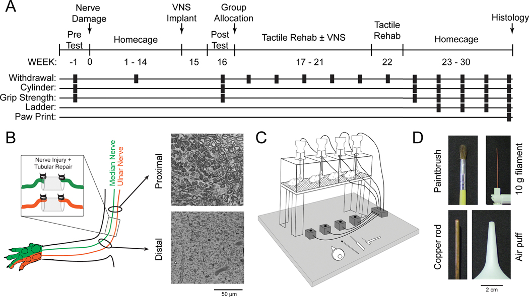
Fig. 1. Experimental Design and Tactile Rehabilitation Paradigm.
(A) Timeline of experimental design illustrating when each assessment is performed. (B) Schematic and representative images from proximal and distal cross-sections of the median nerve approximately 30 weeks after nerve transection and tubular repair. Reinnervation takes place, but the procedure results in chronic deficits in nerve architecture distal to the injury site. (C) Schematic of the tactile rehabilitation apparatus. Rats were placed in individual cages with a wire mesh floor. A variety of tactile stimuli were applied to the ventral surface of the right (injured) forepaw. A button press coincident with the delivery of the tactile stimuli initiated a 500 ms train of VNS in the appropriate group. (D) Detailed view of the devices utilized during tactile rehabilitation. The stimuli were selected to encompass a wide range of somatosensory features.

