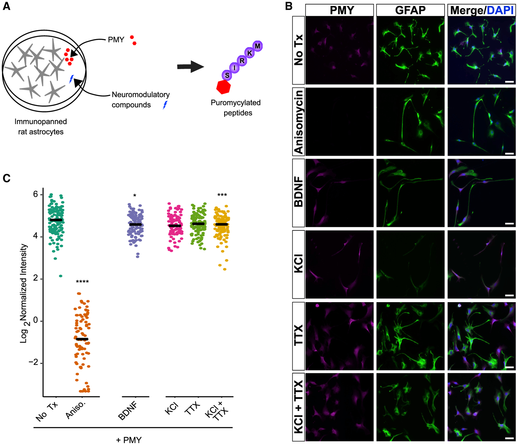
Figure 4. The effect of BDNF and KCl on astrocyte translation requires neurons.

(A) Schematic of the assay for astrocyte translation. Purified astrocytes are treated with test compounds, and the resulting translational response is measured using PMY, which tags nascent peptides and is visualized for the subsequent immunofluorescence.
(B) Representative fields of immunopanned astrocytes, stained for PMY and GFAP. Scale bar, 50 μm.
(C) Quantification of PMY intensity in astrocytes. Mean intensity (signal/area) was calculated for individual cells. Pairwise t tests were performed compared with no Tx. ****p < 0.001, ***p < 0.005, *p < 0.05. Ncells (condition) = 129 (no Tx; i.e., PMY only), 87 (anisomycin), 97 (KCl), 120 (TTX), 104 (BDNF), 111 (KCl + TTX).
