Figure 3.
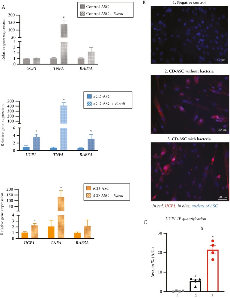
E. coli promotes browning in adipose stem cells isolated from creeping fat. [A] Relative gene expression analysis of the browning marker UCP1, the inflammatory marker TNFA and the phagocytic marker RAB5A in adipose stem cells [ASCs] isolated from visceral adipose tissue [VAT] of patients with active or inactive Crohn’s disease [CD] or from healthy controls, co-cultured or not for 3 h with E. coli. Bars in graphs represent mean ± SEM and significant differences vs cells without E. coli [*p < 0.05] [n = 3 per group]. [B] Immunofluorescence of UCP1 [red label] in ASCs isolated from patients with active CD co-cultured or not with E. coli [3 h]. DAPI staining of nuclei in blue. *p < 0.05 vs control [n = 4 per group]. [C] UCP1 immunofluorescence quantification [% of fluorescence area, mean values ± SEM] [n = 4 per group].

