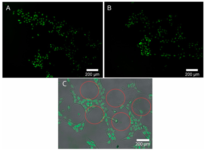
Figure 4.
(A) Adhered hiPSC cells in the pattern printed on the culture plate. (B) Cells attached to a pattern of different heights with higher cell densities after 12 h of incubation. (C) Brightfield and overlay layer that show how the cells follow only the segment where the pattern was printed. Red outlines show the dots of the patterns created by the microcontact printing technique; the cells grew around the dots.

