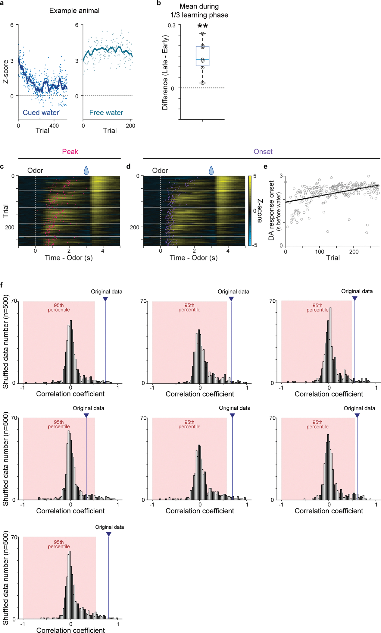
Extended Data Fig. 2. Dopamine release in the ventral striatum during first-time classical conditioning.

(a) The time-course of dopamine sensor responses to cued water (left) and to free water (right) in an example mouse. Each dot represents responses in each trial, and a line shows moving averages of 20 trials. (b) Dopamine response to reward-associated cue in the late phase (2–3s after cue onset) was significantly higher than activity in the early phase (0–1s after cue onset) during the first 1/3 of learning phase (t =5.0, p = 0.22 × 10⁻²; two-sided t-test). (c) Dopamine sensor signal peaks during delay periods (red) overlaid on a heatmap of dopamine sensor signals in cued water trials. n=7 animals. (d) Dopamine sensor response onset (purple) overlaid on a heatmap of dopamine sensor signals. (e) Linear regression of dopamine excitation onset with trial number. (f) Each panel shows correlation coefficients between activity peak and trial number for trial order shuffled data (n=500) in each animal (7 animals). 95 percentile area is marked with red and the correlation coefficient of original data is shown as blue line. Center of boxplot showing median, edges are 25th and 75th percentile, and whiskers are most extreme data points. **p<0.01.
